Evos FL Microscope
Location: HSC (Building 586) Room 54
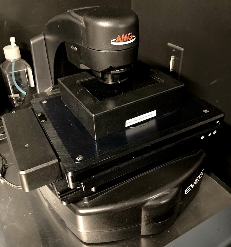
The EVOS FL Imaging system is an all-in-one fluorescence microscope designed for efficiency and ease of use. The on-board computer and integrated imaging software allows users to capture and save four channels fluorescence images/data directly from the microscope. The EVOS FL Imaging System is suitable for checking samples before using a high-end microscope system.
Main features of System:
- Dual 16-bit CCD acquisition cameras (Monochromatic and Color)
- 22” high-resolution touch screen color monitor
- Automated X-Y scanning stage with interchangable specimen holder inserts. We have available inserts for glass slides, culture dishes, multiwell plates, and T-flasks
- EVOS LED Light Cube Technology. We have following cubes available:
DAPI, GFP, RFP, Texas Red, and Cy5
Suggested Applications:
-
Fixed and live cell imaging.
- Tissue imaging.
- Sample check before using a high-end microscope system.
Light Sources:
LED light cubes which combine bright LED illumination with excitation and emission filters. DAPI, CY5, GFP, RFP and Texas Red cubes are available. Four can be installed at one time. Contact Cell Imaging staff to change cubes if needed. A selection guide for matching cubes to dyes is on the ThermoFisher site: Guide
| Cube | Excitation (nm) | Emission (nm) |
| DAPI | 357/44 | 447/60 |
| GFP | 485/25 | 524/24 |
| RFP | 531/40 | 593/40 |
| Texas Red | 585/29 | 628/32 |
| Cy5 | 628/40 | 692/40 |
Objectives:
- objectives on the Evos are Long Working Distance, not Coverslip-Corrected. They are optimized for use through vessels with a nominal wall thickness of 0.9-1.5 mm.
Objective Magnification Immersion Numerical
Apperature
Correction Ring Coverglass (mm) Working
Distance (mm)
Link PlanApo 1.25X Air 0.04 5.10
Plan Fluor 4X Air 0.13 17.2
Plan Fluor 10X Air 0.3 1.2 8.31 Plan Fluor 20X Air 0.45 1.2 7.11 Plan Fluor 40X airAir 0.65 1.2 2.81
The