Instrument Details
Details about each instrument
- Zeiss 700 Confocal Microscope
- Prairie Ultima 2
- Nikon A1R Confocal
- Evos Auto Color and Fluor Microscope
- Zeiss Axio Scan.Z1
- Nikon Automated Widefield Microscope
- Leica SP8 White Light System
- Leica SP8 405-488-561-633 Laser Confocal
- Zeiss Axioscan 7
- Zeiss 880 Airy Scan
- StedyCon
- Olympus FV1000 Confocal Microscope
- Delta Vision Widefield
- Leica Spinning Disk Confocal
- Nikon Ring TIRF or Spinning Disk Confocal
Zeiss 700 Confocal Microscope
Location: HSC (Building 586) Room 56
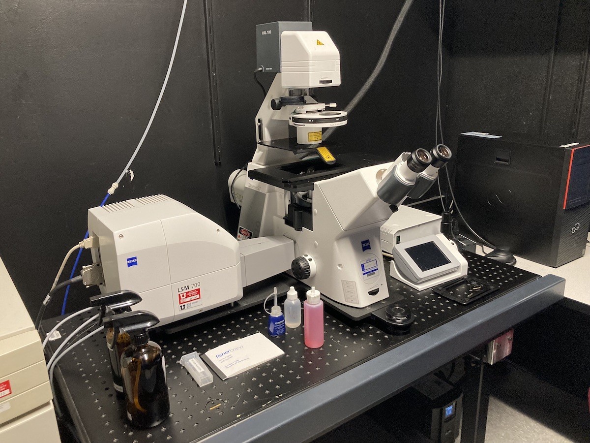
The LSM 700 laser scanning confocal microscope is the seventh generation of confocal microscopes from Carl Zeiss. The LSM 700 system uses a Zeiss AXIO Observer Z1 inverted microscope with transmitted illumination (HAL 100) and a laser illumination source. The LSM 700 system features easy operation, excellent sensitivity, and a design that can handle complex tasks.
Main features of Zeiss 700 System:
- Two Photo Multipliers Tube (PMT1 and PMT2) detector unit enables simultaneous 12 bit (4096 intensity levels) acquisition of up to 4 fluorescent channels.
- Confocal scan head unit can provide high-resolution images up to 2048 x 2048 pixels in size.
- Fully automated Zeiss Observer Z1 inverted microscope.
- LED Epifluorescence and HAL100 Brightfield illumination lamps for rapid sample identification and focusing through the eyepiece.
- Filter wheel used for epifluorescence viewing of the sample is currently outfitted with DAPI , FITC (green), and Rhodamine (red) filter cube sets.
- Motorized XY stage with a variety of stage inserts available (slides, dishes, multiwall plates, etc..).
- Zeiss Zen Black multi-platform acquisition software.
- Measurements include: Z stack, Time lapse, Stitching/Tiling, XY positions, and Spectra unmixing
Suggested Applications:
-
3D Imaging.
- Multichannel fluorescence fixed slide imaging.
- Tiled mosaics.
Imaging lasers:
| Wavelength (nm) | Type | Power | Manufacturer |
| 405 | diode | 5 mW |
Lasos Lasertechnik GmbH |
| 488 | diode | 10 mW |
Lasos Lasertechnik GmbH |
| 555 | diode | 10 mW |
Lasos Lasertechnik GmbH |
| 639 | diode | 5 mW |
Lasos Lasertechnik GmbH |
Objectives:
| Objective | Magnification | Immersion |
Numerical Apperature |
Correction Ring | Coverglass (mm) |
Working Distance (mm) |
| EC Plan Neofluar M27 | 10X | Air | 0.3 | 0.17 |
5.2 |
|
| Pan Apo M27 | 20X | Air | 0.8 | 0.17 | 0.55 | |
| LD C-Apo Korr M27 | 40X | Water | 1.1 | Corr | 0.14-0.19 | 0.62 |
| Pan Apo DIC M27 | 63X | Oil | 1.4 | 0.17 | 0.19 |
Prairie Ultima 2
Location: HSC Room 48B / 48D
Main features of Prairie Ultima 2/Multi-Photon Confocal System:
- Six detectors, four reflected mode Photo Multipliers Tube (PMT) detectors and two transmitted mode PMTs for second harmonic and fluorescence detection.
- High NA water immersion optics including 16x PLANAPO LWD Nikon NA 0.8 and a PLANAPO Correction collar LWD 25x NA 1.1, 60x LUMFL water immersion Olympus NA0.8.
- Resonance scanner for high speed imaging timelapse (~200fps depending on format)
- Piezo Z drive for fast z acquisition, 2 inch travel, XY motorized stage
- Fixed (Z) stage with adjustable (2 inch) travel
- Bioscience Tools stage top incubator with heated pad, heated dish
- Mouse Anesthesia system from VetEquip for in vivo applications (Halothane with scrubber)
- Gas supply, 02, C02, 02/C02 mix
Imaging lasers:
- Coherent Vision II with tuning 705-1060nm pulsed femtosecond range 4000-400mW depending on wavelength
Suggested Applications:
- Deep live cell imaging of zebrafish, mouse in vivo, organ culture and slices, 3D cell culture
- Multichannel fluorescence in vivo, slices or cultures
- Tiling mosaics of fixed or live samples
- Timelapse of whole mount, in vivo mouse, culture
- Second harmonic imaging of collagen or other ECM
Objectives:
48B:
| Objective | Magnification | Immersion | Numerical Aperture | Correction Collar | Coverslip (mm) | Working Distance (mm) |
| Apo | 25 | Water, Water Dipping | 1.1 | Corr | 0-0.17 | 2 |
48D:
| Objective | Magnification | Immersion | Numerical Aperture | Correction Collar | Coverslip (mm) | Working Distance (mm) |
| Plan Apo Lambda | 4 | Air | 0.2 |
20 |
||
| Plan Fluor | 10 | Air | 0.3 | 0.17 |
16 |
|
| UPlanFl | 10 | Air | 0.3 |
|
||
| LWD | 16 | Water Dipping | 0.8 | 0 |
3 |
|
| Apo | 25 | Water, Water Dipping | 1.1 | Corr | 0-0.17 |
2 |
| Fluor | 40 | Water Dipping | 0.8 | 0 |
2 |
Nikon A1R Confocal
Location: HSC Room 56
Main features of System:
- Four Photo Multipliers Tube (PMT) detector unit enables simultaneous 12 bit (4096 intensity levels) acquisition of up to 4 fluorescent channels.
- Fifth transmitted light detector available for simultaneous brightfield imaging of samples while scanning confocally.
- Hybrid confocal scan head unit can provide high resolution images up to 4096 x 4096 pixels in size in conventional galvonometer mode and in high speed resonant mode up to 30 frames per second at 512 x 512 pixels.
- Ultra high speed resonant acquisitions up to 420 frames per second at 512 x 32 pixels.
- Fully automated Nikon Ti-E inverted microscope.
- Epifluorescence and DIC (Nomarski) illumination lamps for rapid sample identification and focusing.
- Filter wheel used for epifluorescence viewing of sample is currently outfitted with with DAPI (blue), FITC (green), and TRITC (red), filter cube sets.
- ASI MS-2000 Motorized XY stage with a variety of stage inserts available (slides,dishes, multiwall plates, etc..).
- MCL Piezo Stage (100um range) for high speed 3D (i.e., Z-stack) volume acquisition.
- Perfect Focus System provides focal depth consistency across varying sample regions of interest.
- Nikon NIS-Elements multi-platform acquisition software.
Imaging lasers:
- 405nm diode laser
- 488nm Argon gas laser
- 561nm Sapphire diode laser
- 638nm diode laser
Suggested Applications:
- Multichannel fluorescence and transmitted fixed slide imaging
- Tiling mosaics of fixed or live samples
- Timelapse of dishes or glass bottom well plates
Objectives:
| Objective | Magnification | Immersion | Numerical Aperture | Correction Ring | Coverglass | Working Distance |
| Plan Apo Lambda | 4 | Air | 0.2 | 20 | ||
| Plan Apo Lambda | 10 | Air | 0.45 | 0.17 | 4 | |
| Plan Apo Lambda | 20 | Air | 0.75 | 0.17 | 1 | |
| Plan Fluor | 40 | Oil | 1.3 | 0.17 | 0.2 | |
| Plan Apo Lambda | 60 | Oil | 1.4 | 0.17 | 0.13 |
Other objectives available. Please inquire with Core personnel
Evos Auto Color and Fluor Microscope
Location: HSC (Building 586) Room 54
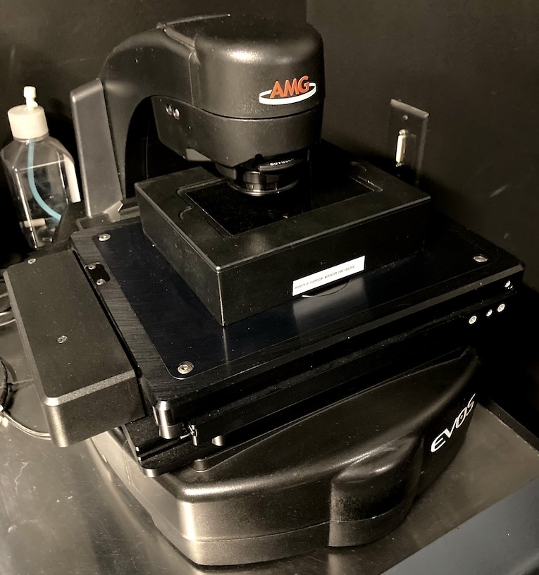
The EVOS FL Imaging system is an all-in-one fluorescence microscope designed for efficiency and ease of use. The on-board computer and integrated imaging software allows users to capture and save four channels fluorescence images/data directly from the microscope. The EVOS FL Imaging System is suitable for checking samples before using a high-end microscope system.
Main features of System:
- Dual 16-bit CCD acquisition cameras (Monochromatic and Color)
- 22” high-resolution touch screen color monitor
- Automated X-Y scanning stage with interchangable specimen holder inserts. We have available inserts for glass slides, culture dishes, multiwell plates, and T-flasks
- EVOS LED Light Cube Technology. We have following cubes available:
DAPI, GFP, RFP, Texas Red, and Cy5
Suggested Applications:
-
Fixed and live cell imaging.
- Tissue imaging.
- Sample check before using a high-end microscope system.
Light Sources:
LED light cubes which combine bright LED illumination with excitation and emission filters. DAPI, CY5, GFP, RFP and Texas Red cubes are available. Four can be installed at one time. Contact Cell Imaging staff to change cubes if needed. A selection guide for matching cubes to dyes is on the ThermoFisher site: Guide
| Cube | Excitation (nm) | Emission (nm) |
| DAPI | 357/44 | 447/60 |
| GFP | 485/25 | 524/24 |
| RFP | 531/40 | 593/40 |
| Texas Red | 585/29 | 628/32 |
| Cy5 | 628/40 | 692/40 |
Objectives:
The objectives on the Evos are Long Working Distance, not Coverslip-Corrected. They are optimized for use through vessels with a nominal wall thickness of 0.9-1.5 mm.
| Objective | Magnification | Immersion |
Numerical Apperature |
Correction Ring | Coverglass (mm) |
Working Distance (mm) |
| PlanApo | 1.25X | Air | 0.04 |
5.10 |
||
| Plan Fluor | 4X | Air | 0.13 |
17.2 |
||
| Plan Fluor | 10X | Air | 0.3 | 1.2 | 8.31 | |
| Plan Fluor | 20X | Air | 0.45 | 1.2 | 7.11 | |
| Plan Fluor | 40X | Air | 0.65 | 1.2 | 2.81 |
Zeiss Axio Scan.Z1
Location: HSC (Building 586) Room 60
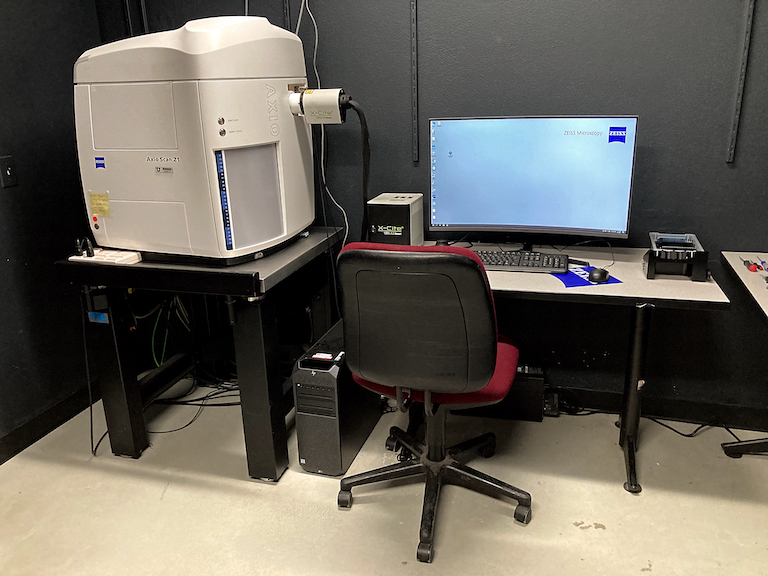
The Zeiss Axio-Scan.Z1 is a fast and flexible slide scanner that digitizes specimens to produce high-quality virtual slides. In addition to brightfield slide scanning, the Axio-Scan.Z1 uses LED lights to scan up to nine fluorescent channels. Up to 100 slides can be loaded onto the scanner, making it suitable for large projects, including digital archives. Scanned images are saved as CZI files and can be viewed using Zeiss freeware Zen-lite or third-party image analysis programs.
Main features of Axio Scan.Z1:
- Holds up to 100 slides.
- Able to image both bright-field (e.g. H&E slides) and up to nine-colour fluorescent samples.
- Image areas can be detected automatically or marked out, to form montages of the whole sample, or multiple samples on each slide
- Setup wizard to guide users through optimising image acquisition and autofocus parameters.
- Once setup, the system can be left for unattended automated scanning e.g. overnight
Light Sources:
In addition to an internal lamp for brightfield imaging, the Axio Scan.Z1 has an X-Cite 120LED source for fluorescent imaging.
Filter cubes for LEDs:
The scanner has a 10 position filter turret to fit most fluorophores. The Zen profile wizard is used to pick filter to match your imaging needs. Alternate filter cubes can be installed for specialty applications. Please consult with Cell Imaging staff for consultation and assistance.
| Filter Position | Excitation (nm) | Emission (nm) | Typical Fluorophores | Opal Dye |
| 1 | empty | empty | Brightfield | |
| 2 | 325-375 | 435-485 | DAPI | |
| 3 | 426-446 | 465-495 | Cyan, Protein | 480 |
| 4 | 480-500 | 510-530 | Alexa Fluor 488 | 520 |
| 5 | 510-530 | 540-560 | Alexa Fluor 489 | 540 |
| 6 | 538-562 | 570-640 | HE, DsRed, Alexa Fluor 555 | 570 |
| 7 | 568-592 | 610-640 | Alexa Fluor 568 | 620 |
| 8 | 610-630 | 645-675 | Alexa Fluor 660 | 650 |
| 9 | 625-655 | 666-715 | Cy5 | |
| 10 | 650-680 | 695-755 | Alexa Fluor 680 | 690 |
Objectives:
| Objective | Magnification | Immersion |
Numerical Apperature |
Correction Ring | Coverglass (mm) |
Working Distance (mm) |
| EC Plan-Neofluar M27 | 2.5X | Air |
0.085 |
0.17 |
8.8 |
|
| Fluor M27 | 2.5X | Air |
0.12 |
0.17 |
8.7 |
|
| Fluar M27 | 5X | Air | 0.25 | 0.17 |
12.5 |
|
| Plan Apo M27 | 10X | Air | 0.45 | 0.17 |
2.1 |
|
| Plan Apo M27 | 20X | Air | 0.8 | 0.17 | 0.55 | |
| Plan Apo Korr M27 | 40X | Air | 0.95 | 0.13-0.21 | 0.25 |
Nikon Automated Widefield Microscope
Location: HCI Room 1430
Main features of System:
- Widefield light pathway employs a high sensitivity Andor Zyla CMOS camera
- Sola automated LED lightsource for both widefield fluorescence and transmitted illumination pathways
- Fully automated Nikon Ti-E inverted microscope
- Perfect Focus System provides focal depth consistency across varying sample regions of interest
- Motorized Prior II XY stage with a variety of stage inserts available (slides,dishes, multiwall plates, etc..)
- Nikon NIS-Elements multi-platform acquisition software capable of Tiling, timelapse, scripting, analysis and advanced image processing.
- Stage Incubator: OKOLAB, C02 and Humidity with either dish or plate adaptor
Suggested Applications:
- Multichannel fluorescence and transmitted fixed slide imaging
- Tiling mosaics of fixed or live samples
- Timelapse of dishes or glass bottom well plates
Objectives:
| Objective | Magnification | Immersion | Numerical Aperture | Correction Ring | Coverglass (mm) | Working Distance (mm) |
| Plan Apo Lambda | 4 | Air | 0.2 | 20 | ||
| Plan Apo Lambda | 10 | Air | 0.45 |
0.17 |
4 | |
| Plan Apo Lambda | 20 | Air | 0.75 | 0.17 | 1 | |
| S PLAN Fluor ADM ELWD | 20 | Air | 0.45 | Corr | 0-2.0 | 8.2-6.9 |
| Plan Apo Lambda | 60 | Oil | 1.4 | 0.17 | 0.13 | |
| Plan Apo Lambda | 100 | Oil | 1.45 | 0.17 | 0.13 |
Other objectives available. Please inquire with Core personnel
Leica SP8 White Light System
Location: HCI Room 1480
Main features of Leica SP8 White Light System:
- Five detectors, Two Photo Multipliers Tube (PMT) detectors and three HyD detectors for enhanced sensitivity and dynamic range for 5 total fluorescent channels.
- Transmitted light detector available for simultaneous brightfield imaging of samples while scanning confocally.
- Hybrid confocal scan head unit can provide high resolution images up to 4096 x 4096 pixels in size in conventional galvonometer mode and in high speed resonant mode up to 30 frames per second at 512 x 512 pixels.
- Ultra high speed resonant acquisitions up to 420 frames per second at 512 x 32 pixels.
- Fully automated Leica DM8 microscope
- Epifluorescence and DIC (Nomarski) illumination lamps for rapid sample identification and focusing.
- Filter wheel used for epifluorescence viewing of sample is currently outfitted with with DAPI (blue), FITC (green), and TRITC (red), filter cube sets.
- Laser autofocus system provides focal depth consistency across varying sample regions of interest.
- OkoLab heating/cooling stage top incubator with C02 and humidity. Plates and dishes with glass bottoms.
Imaging lasers:
- 405nm diode laser
- 488nm Argon gas laser
- White Light continuously tunable pulsed laser 440-700nm
Suggested Applications:
- Multichannel fluorescence and transmitted fixed slide imaging
- Tiling mosaics of fixed or live samples
- Timelapse of dishes or glass bottom well plates
Objectives:
| Objective | Magnification | Immersion | Numerical Aperture | Correction Ring | Coverglass (mm) | Working Distance (mm) |
| HC PL APO CS2 | 10 | Air | 0.4 | 2.74 | ||
| HC PL APO CS2 | 20 | Either water, glycerine, or oil | 0.75 | Corr | 0.66 | |
| HC PL APO CS2 | 20 | Air | 0.75 | 0.17 | 0.62 | |
| HC PL APO CS2 | 40 | Water | 1.1 | Corr | 0.14-0.18 | 0.65 |
| HC PL APO CS2 | 40 | Oil | 1.3 | 0.17 | 0.24 | |
| HC PL APO CS2 | 60 | Oil | 1.4 | 0.17 | 0.14 |
Other objectives available. Please inquire with Core personnel
Leica SP8 405-488-561-633 Laser Confocal
Location: HCI Room 1440
Main features of Leica SP8 White Light System:
- Five detectors, Two Photo Multipliers Tube (PMT) detectors and three HyD detectors for enhanced sensitivity and dynamic range for 5 total fluorescent channels.
- Transmitted light detector available for simultaneous brightfield imaging of samples while scanning confocally.
- Hybrid confocal scan head unit can provide high resolution images up to 4096 x 4096 pixels in size in conventional galvonometer mode and in high speed resonant mode up to 30 frames per second at 512 x 512 pixels.
- Ultra high speed resonant acquisitions up to 420 frames per second at 512 x 32 pixels.
- Fully automated Leica DM8 microscope
- Epifluorescence and DIC (Nomarski) illumination lamps for rapid sample identification and focusing.
- Filter wheel used for epifluorescence viewing of sample is currently outfitted with with DAPI (blue), FITC (green), and TRITC (red), filter cube sets.
- Laser autofocus system provides focal depth consistency across varying sample regions of interest.
- OkoLab heating/cooling stage top incubator with C02 and humidity. Plates and dishes with glass bottoms.
- z-galvo
Imaging lasers:
- 405nm diode laser
- 488nm Argon gas laser
- White Light continuously tunable pulsed laser 440-700nm
- 561
- 633
Suggested Applications:
- Multichannel fluorescence and transmitted fixed slide imaging
- Tiling mosaics of fixed or live samples
- Timelapse of dishes or glass bottom well plates
Objectives:
| Objective | Magnification | Immersion | Numerical Aperture | Correction Collar | Coverslip (mm) | Working Distance (mm) |
| HC PL APO CS2 | 10 | Air | 0.4 | 2.74 | ||
| HC PL APO CS2 | 20 | Either water, glycerine, or oil | 0.75 | Corr | 0.66 | |
| HC PL APO CS2 | 20 | Air | 0.75 | 0.17 | 0.62 | |
| HC PL APO CS2 | 40 | Water | 1.1 | Corr | 0.14-0.18 | 0.65 |
| HC PL APO CS2 | 40 | Oil | 1.3 | 0.17 | 0.24 | |
| HC PL APO CS2 | 63 | Oil | 1.4 | 0.17 | 0.14 |
Zeiss Axioscan 7
Location: HCI Room 1470

The Zeiss AxioScan 7 is a fast and flexible slide scanner that digitizes specimens to produce high-quality virtual slides. In addition to brightfield slide scanning, the Axioscan 7 can scan up to five fluorescent channels using fast switching LED lights. Up to 100 slides can be loaded onto the scanner, making it suitable for large projects, including digital archives. Scanned images are saved as CZI files and can be viewed using Zeiss freeware Zen-lite or third-party image analysis programs.
Main Features of the Axioscan 7:
- Holds up to 100 slides.
- Able to image both bright-field (e.g. H&E slides) and up to nine-colour fluorescent samples.
- Image areas can be detected automatically or marked out, to form montages of the whole sample, or multiple samples on each slide
- Setup wizard to guide users through optimising image acquisition and autofocus parameters.
- Once setup, the system can be left for unattended automated scanning e.g. overnight
Suggested Applications:
- Fixed slides.
Configurations:
The Axioscan 7 has a replaceable filter turret and can be switched between two light sources. The 10 position filter cube turret is typically used with the Colibri 7 switchable LED source. The fast filter wheel is typically used with the X-Cite Xylis LED source.
| Configuration | Light Source | Filter Set |
| 1 | Colibri 7 | Filter Cubes |
| 2 | X-Cite Xylis | Fast Filter Wheel |
Only Cell Imaging staff are permitted to switch between configurations. Please schedule our time and plan accordingly.
Light Sources:
Colibri 7:
The Colibri 7 has 6 LEDs with excitation filters to provide 7 excitation bands. (One LED has a motorized filter to split it into two bands.)
Excitation Bands (There is one too many here)
| Band | Wavelength / Bandwidth (nm) | Some typical fluorophores |
| UV | 385/30 |
API, Hoechst 33342, Hoechst 33258, Alexa Fluor 350, Alexa Fluor 405, Indo-1, eBFP / BFP, eGFP (wt), True Blue |
| V | 423/44 |
Pacific Blue, Lucifer Yellow, Alexa Fluor 433, eCFP, Cerulean |
| B | 469/38 |
FM1-43, Cy2, eGFP, NBD, MitoTracker Green, Alexa Fluor 488, BCECF, Calcein, DiO SNAFL, YO-Pro-1, Nissl, LysoSensor Green, mHoneydew, FITC / Fluorescein, Kaede (green / red), PerCP, YoYo-1,FuraRed |
| C | 511/44 |
Rhodamine 123, Fluo-4, Oregon Green BAPTA, Sytox Green, eYFP, FM4-64, Eosin/HE, Acridine Orange, JC1, Bodipy FL, Propidium Iodide, Spectrum Green, Calcium Green |
| G | 555/30 |
TRITC, 7-AAD, Cy3, tdTomato, Alexa Fluor 546, Alexa Fluor 555, DsRed, mOrange, TagRFP, SNARF, DyLight 549, Spectrum Orange |
| Y | 590/27 |
MitoTracker RED FM/CMXRos, txRed, mCherry, mRFP1, Cy3.5, Rhodamine B, Alexa Fluor 568, Dylight 594, Alexa Fluor 594, Bodipy TR |
| R | 631/33 |
Alexa Fluor 633, Alexa Fluor 647, Cy5, DRAQ5, ToTo-3, ATTO-655, MitoTracker DeepRed, APC, ATTO-647N |
| FR | 735/40 |
Alexa Fluor 750, Alexa Fluor 790, Cy7, Cy7.5 |
X-Cite Xylis:
The X-Cite Xylis XT720L is a broad spectrum illuminator with a range from 380-770nm. It has peaks at 385, 430, 475, 545, 635, 735nm.
Filters:
Filter Cubes:
In multi-band fluorescence imaging, the filter turret has to move between positions for each band. This is usually done on a per-tile basis. At about 400ms per move, this can add up, especially for a large scan at higher magnification. The Axioscan 7 has 2 multi-band cubes. If these can be used, it can reduce or eliminate these moves and reduce scan time. These can be used on the Axioscan 7 because it has the Colibri 7 switching LED source.
|
Filter No |
Fluorochrome |
Excitation (nm) |
Beamsplitter (nm) |
Emission (nm) |
|
1 |
Empty (Brightfield) |
|
|
|
|
2 |
HE GFP |
450-490 |
495 |
500-550 |
|
3 |
HE DsRed |
538-562 |
570 |
570-640 |
|
4 |
Cy5 |
625-655 |
660 |
665-715 |
|
5 |
HE BFP |
370-410 |
420 |
430-470 |
|
6 |
HE DAPI/GFP/Cy3.5/Cy7 |
375-395 455-483 583-600 720-750 |
405 493 611 762 |
410-440 501-547 618-650 770-800 |
|
7 |
HE DAPI/GFP/Cy3.5/Cy5/Cy7 |
370-400 450-488 540-570 614-647 720-750 |
405 493 575 654 761 |
412-438 501-527 582-601 662-700 770-800 |
|
Pol |
Circular polarizer and analyzer |
|
|
|
|
ND |
6% Neutral Density Filter |
|
|
|
Fast Filter Wheel:
The fast filter wheel moves between filter positions about 10 times faster than the filter cube turret.
Need information here
| Filter | Pass Peak and Width | Typical fluorophores |
| Empty (Brightfield) | ||
| Single Channel 1 | ||
| Single Channel 2 | ||
| Single Channel 3 | ||
| Dual Channel 4 | ||
| Dual Channel 5 |
Objectives:
| Objective | Magnification | Immersion |
Numerical Apperature |
Correction Ring | Coverglass (mm) |
Working Distance (mm) |
| Fluar M27 | 5X | Air | 0.25 | 0.17 |
12.5 |
|
| Pan Apo M27 | 10X | Air | 0.45 | 0.17 | 2.1 | |
| Pan Apo M27 | 20X | Air | 0.8 | 0.17 | 0.55 | |
| Pan Apo Korr M27 | 40X | Air | 0.95 | 0.13-0.21 | 0.25 | |
| EC Pan-Neofluar Pol M27 | 20X | Air | 0.5 | 0.17 | 2 |
Zeiss 880 Airy Scan
Location:CSC Room 032
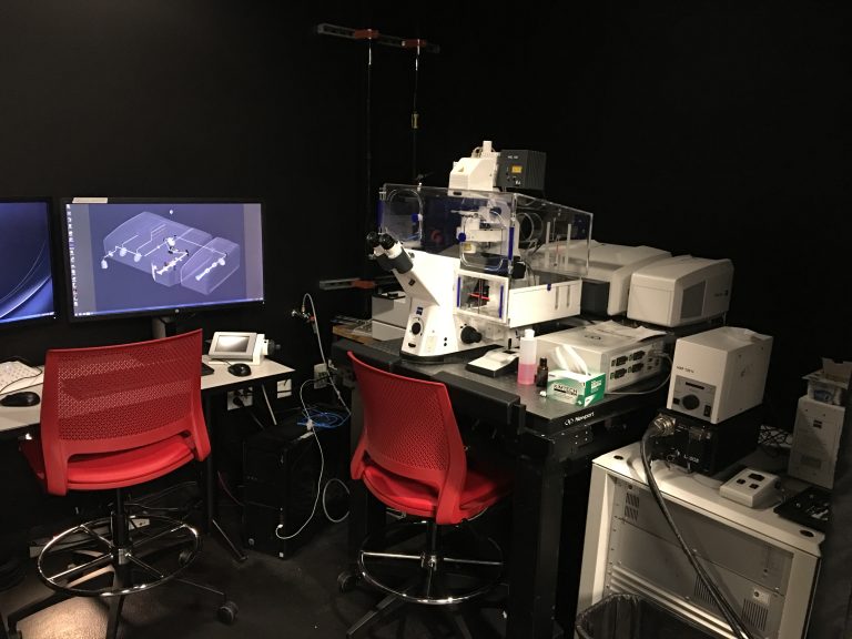
Main features of Zeiss 880 Airy Scan System:
- Five detectors, Three Photo Multipliers Tube (PMT) detectors and two GaSP detectors for enhanced sensitivity and dynamic range for 5 total fluorescent channels.
- Transmitted light detector available for simultaneous brightfield imaging of samples while scanning confocally.
- AIRY scan detector capable of super resolution imaging
- Fast AIRY scan system for high speed super resolution imaging timelapse
- Epifluorescence and DIC (Nomarski) illumination lamps for rapid sample identification and focusing.
- Filter wheel used for epifluorescence viewing of sample is currently outfitted with with DAPI (blue), FITC (green), and TRITC (red), filter cube sets.
- Laser autofocus system provides focal depth consistency across varying sample regions of interest.
- Piezo Z stage for fast z acquisition
- IBIDI stage top incubator with temperature, C02 and humidity. Plates and dishes with glass bottoms.
Imaging lasers:
- 405nm diode laser
- 488nm Argon gas laser
- 561nm diode
- 638nm diode
Suggested Applications:
- Multichannel fluorescence and transmitted fixed slide imaging
- Tiling mosaics of fixed or live samples
- Timelapse of dishes or glass bottom well plates
Objectives:
|
Objective |
Magnification | Immersion | Numerical Apperature | Correction Ring | Coverglass (mm) | Working Distance (mm) |
| Plan Apo M27 | 5 | Air | 0.16 | 0.17 | 12.1 | |
| EC Plan-Neofluar M27 | 10 | Air | 0.3 | 0.17 | 5.2 | |
| Plan Apo M27 | 20 | Air | 0.8 | 0.17 | 0.55 | |
| Plan Apo M27 | 40 | Oil | 1.3 | 0.17 | 0.21 | |
| Plan Apo DIC M27 | 63 | Oil | 1.4 | 0.17 | 0.19 | |
| Calibration Apo DIC M27 |
StedyCon
Location: CSC Room 30
Main features of StedyCon:
- STEDYCON from Abberior is a new class of nanoscope with multicolor confocal and STED (Stimulated emission depletion) capabilities. The lateral STED resolution could reach 30nm depending on dyes and objectives used. The acquired STEDYCON system is fitted on a Nikon Ti-S-E inverted microscope from a camera port and equipped with four excitation lasers: 405 nm CW and 488, 561, 640 nm pulse laser.
- The STED process is applied by a 775 nm pulse laser. Four highly sensitive single-photon-counting Avalanche photodiode detectors (APD) are mounted.
- Two STED and four confocal imaging channels can be detected. A 100X/NA=1.45 oil immersion lens and other low magnification lens are provided to satisfy versatile measurements.
- An XY stage from Marzhauser and an ultra-fast and precise Piezo Z stage from PIFOC are mounted. Browser-based acquisition software is installed on a powerful PC with a Windows 10 system.
- Other advanced functions including Z stack, multipositioning, time series, and tiling and stitching can be performed.
Objectives:
| Objective | Magnification | Immersion | Numerical Apperature | Correction Ring | Coverglass (mm) | Working Distance (mm) |
| Plan Apo Lambda | 4X | Air | 0.2 | 20 | ||
| Okan-Neofluar | 5X | Air | 0.15 | 0.17 | 13.6 | |
| Plan Apo | 10X | Air | 0.45 | 0.17 | 4 | |
| PlanApo Lambda | 20X | Air | 0.75 | 0.17 | 1 | |
| Plan Apo Lambda | 60X | Oil | 1.4 | 0.17 | 0.13 | |
| Plan Apo Lambda | 100x | Oil | 1.45 | 0.17 | 0.13 |
Olympus FV1000 Confocal Microscope
Location: CSC Room 32
Main features of System:
- Three standard Photo Multipliers Tube (PMT) light detectors with two of the channels having tunable spectral bandwidths.
- Simultaneous transmitted light or DIC imaging while scanning confocally.
- Fully automated Olympus IX81 inverted microscope.
- Additional epifluorescence and DIC (Nomarski) illumination lamps for rapid sample identification and focusing.
- Motorized Prior II XY stage with a variety of stage inserts available (slides,dishes, multiwall plates, etc..).
- Olympus FV10-ASW 3.1 confocal imaging software
Imaging lasers:
- 405nm diode laser
- 488nm Argon gas laser
- 543nm Helium-neon gas laser
- 633nm Helium-neon gas laser
Objectives:
| Objective | Magnification | Immersion | Numerical Apperature | Correction Ring | Coverglass (mm) | Working Distance (mm) |
| UPlanSApo | 10X | Air | 0.4 | 0.17 | 3.1 | |
| UPlanApo | 20X | Air | 0.7 | 0.17 | 0.65 | |
| UApo | 40X | Oil | 1.35 | Corr | 0.17 | 0.1 |
| PlanApo | 60X | Water | 1.2 | Corr | 0.13-0.21 | 0.28 |
| PlanApo | 60X | Oil | 1.4 | 0.17 | 0.12 |
Delta Vision Widefield
Location: EEJMRB Room 5100

Hardware:
- Customized Olympus IX73 microscope base w/ TruLight Illuminator
- Olympus X-line PlanApo Objectives 4x, 10x, 20x Dry and 60x Oil
- Ultimate Focus laser-based autofocus system
- Custom XYZ Stage 106mm x 70mm x 7mm
- sCMOS Camera 2040 x 2040 pixels
- 7-channel solid state illuminator
- DIC Transmitted Light Imaging
- Environmental Chamber
Primary applications:
- 2D and 3D Multi-Channel Fluorescence Imaging
- 3D Restorative Deconvolution
- Time-Lapse Imaging
- Multipoint Imaging
Objectives:
| Objective | Magnification | Immersion | Numerical Aperture | Correction Ring | Coverslip (mm) | Working Distance (mm) |
| UPlanXApo | 4 | Air | 0.16 | 13 | ||
| UPlanXApo | 10 | Air | 0.4 | 0.17 |
3.1 |
|
| UPlanXApo | 20 | Air | 0.8 | 0.17 |
0.6 |
|
| UPlanXApo | 40 | Oil | 1.4 | 0.17 |
0.12 |
|
| UPlanXApo | 60 | Oil | 1.42 | 0.17 |
0.15 |
Leica Spinning Disk Confocal
Location: EEJMRB Room 5122
Hardware:
- Leica DMi8 motorized microscope with adaptive focus tracking
- ASI motorized stage with Piezo Z
- W-1 large format spinning disk confocal with beam homogenizer
- Andor 1024 x 1024 EMCCD camera
- Visitron 2D FRAP unit
- 405,488,561,640nm laser lines for DAPI, GFP, RFP, Cy5, and similar fluorochromes
- W-View image splitter for dual channel simultaneous imaging
- LED based widefield fluorescence
- Stage top incubator system
- Visiview acquisition software
Primary applications:
- 2D and 3D Multi-Channel Fluorescence Imaging
- 3D Restorative Deconvolution
- Time-Lapse Imaging
- Multipoint Imaging
- Sample images before (A) and after (B) deconvolution
Objectives:
| Objective | Magnification | Immersion | Numerical Aperture | Correction Ring | Coverglass (mm) | Working Distance (mm) |
| HC PL APO CS2 | 10 | Air | 0.4 | 2.74 | ||
| HCX PL APO CS | 20 | Air | 0.75 | 0.17 | 0.62 | |
| HC PL APO CS2 | 40 | Water | 1.1 | Corr | 0.14-0.18 | 0.65 |
| HC PL APO CS2 | 63 | Oil | 1.4 | 0.17 | 0.14 |
Nikon Ring TIRF or Spinning Disk Confocal
Location: EEJMRB Room 3532
| Objective | Magnification | Immersion | Numerical Apperature | Correction Ring | Coverglass (mm) | Working Distance (mm) |
| Plan Fluor | 10 | Air | 0.3 | 0.17 | 16 | |
| Plan Fluor | 10 | Air | 0.3 | 0.17 | 16 | |
| Plan Apo | 20 | Air | 0.75 | 0.17 | 1 | |
| Apo TIRF | 60 | Oil | 1.49 | Corr | 0.13-0.19 | 0.16-0.07 |
| Plan Apo Lambda | 60 | Oil | 1.49 | 0.17 | 0.13 | |
| Apo TIRF | 100 | Oil | 1.49 | Corr | 0.13-0.19 | 0.16-0.09 |